Herniated Disc Diagnosis
The pain can be most safely eliminated if we know its cause. When we visit a doctor for back pain, we are usually sent for some kind of imaging examination. But what is the difference between them? Below, we have briefly summarized what to expect from each examination and its results.
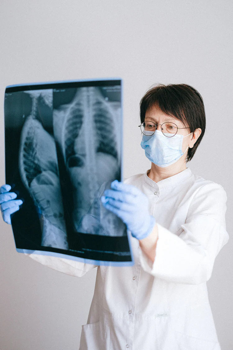
X-ray
What is it suitable for?
It shows the condition of the bones, their positioning, the distance between them (from which the thickness of the intervertebral disc can be inferred), any fractures, cracks, vertebral displacements. Therefore, it is not suitable for determining the condition of the soft tissues, and thus not suitable for detecting herniated discs either.
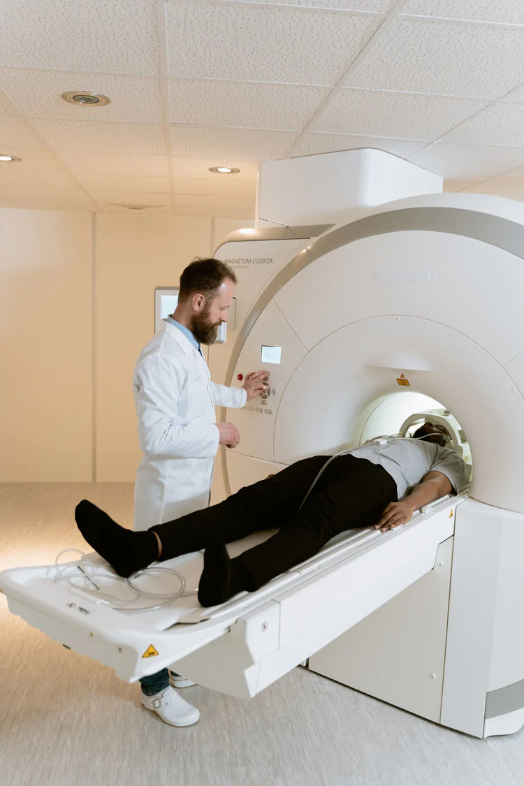
MRI (Magnetic Resonance)
Preparation
The examination is done without X-rays, in a magnetic field. Therefore, no materials containing metal can be brought in, and entry with a pacemaker is prohibited! You have to lie down on a bed, then a magnetic coil is placed on the body. After that, you must remain still.
Examination
The examination takes place inside a tube about 70 cm in diameter, so if someone has a problem with claustrophobia, they should inform before the examination. Help can be requested with a microphone or a signal button. During the examination, a loud buzzing noise can be heard, but earplugs or headphones are used to make this noise more bearable. The examination lasts 20-40 minutes. It is important that in some labs the machines operate with a weight limit of 110-120 kg, so if necessary, inquire about this when scheduling an appointment.
What is it suitable for?
This procedure is already suitable for examining the cervical and thoracic spine
Suitable. It also better shows the soft parts, so nerve roots and disc changes can be detected more clearly. Contrast agents that do not contain iodine can also be used here, which rarely cause allergic reactions but make tumor or inflammatory tissues visible. This examination method can also be used on previously operated spines. It can detect tiny changes that do not even cause complaints for the patient.
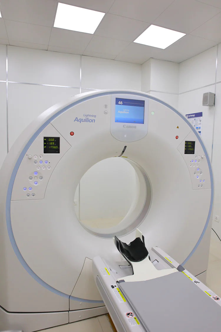
CT (Computer tomography)
Preparation:
It can be requested with a specialist referral and prior appointment. Imaging here is done using X-rays, so children and pregnant women are only examined this way in very justified cases. In some cases, the patient receives a contrast agent intravenously to make the image easier to evaluate. If you have diabetes, heart disease, asthma, or kidney or thyroid conditions, you must always inform the doctor.
Examination:
It is a device similar to a thick ring, with the patient lying on a table at its base. The examination usually lasts 10-15 minutes and causes no pain, except for the intravenous injection. The patient must lie still, occasionally holding their breath, and is informed about this via a microphone. Eating is allowed up to 3-4 hours before the examination, but it is advisable to drink plenty of non-carbonated fluids.
What is it suitable for?
This examination shows not only the bones but also the soft tissues. It is primarily used to examine the lumbar spine. It detects herniated discs, vertebral displacements, fractures, and some tumors as well. The result is usually given on a CD, which must be taken back to the specialist.